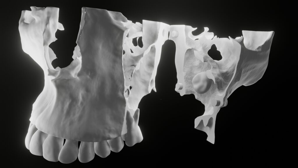
What is Diagnostic 3D Modelling (D3M)?
Ever wondered about focusing solely on the problematic areas, leaving aside unnecessary details? That’s precisely what Diagnostic 3D Modeling (D3M) is all about. Thanks to the latest advancements in 3D design software, especially in the realm of oral radiology, diagnosticians now have the ability to hone in on pathologies by eliminating overlapping structures.
Using D3M, specialists can create a 3D file (STL file) of a specific condition, which can then be easily shared with clinicians or practitioners. These professionals can open the 3D file on their mobile devices or tablets, allowing them to demonstrate and explain the patient’s condition in three dimensions, making it more comprehensible.
This approach not only simplifies patient understanding but also boosts treatment acceptance rates.
What are the advantages of D3M?
Traditional CBCT viewing software must be opened on high-end laptops or desktops as these are graphic-intensive programs. This software requires a learning curve to use efficiently. As most clinicians or practitioners will not have enough time and expertise to use this software, most practitioners advise 2D imaging modalities like OPG.
But with D3M an oral radiologist can send a 3D file, where the clinician, with a single click, can open the 3D file on a regular mobile or Tablet and can show the 3D file directly to the patient. No learning curve is required.
Moreover, the pathology is shown by removing all the superimpositions, where the clinician must just open the image and show a clear pathology to the patient. This facility is not available in conventional CBCT software.
Virtual dissection
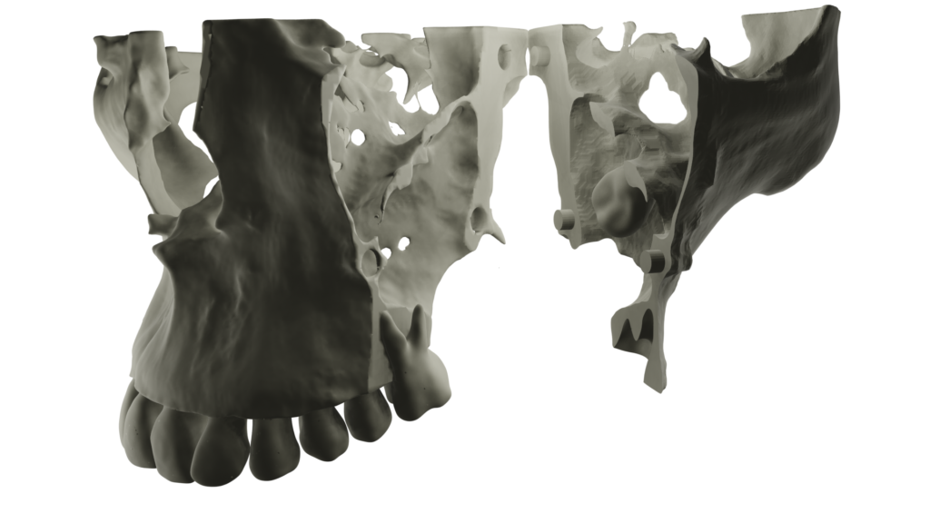
In Diagnostic 3D Models, the incredible capability to virtually dissect a model and explore its internal anatomy offers groundbreaking insights. Take, for instance, a case involving a dentigerous cyst in the maxillary third molar region. By dissecting the maxilla model along an axis intersecting the pathology, we can isolate the dentition from the maxilla itself.
This virtual dissection reveals a wealth of information, including the cystic lining’s impact on repositioning the sinus floor in an upward direction. Furthermore, it unveils the presence of a sizable cystic cavity surrounding the impacted third molar, providing clinicians with a comprehensive and intricate understanding of the condition. This level of detail and precision in diagnostics is truly revolutionary and has the potential to revolutionize patient care in the field of oral health.
3D printing
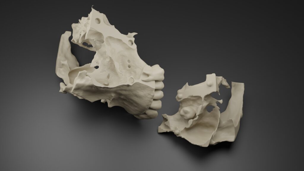
The power of diagnostic 3D modeling goes beyond visualization; it allows for the creation of physical, 3D-printed models that offer unparalleled clarity in depicting intricate anatomical details of pathologies. These printed models serve as invaluable educational tools for patients, enhancing their understanding of their condition and treatment options.
Additionally, surgeons benefit from these models as they can use them for conducting mock surgeries. This hands-on approach provides surgeons with a tangible and precise representation of the patient’s unique anatomy, enabling them to plan and practice procedures with enhanced accuracy. In essence, the combination of 3D modeling and 3D printing is transforming the landscape of patient education and surgical planning, ushering in a new era of comprehensive and patient-centered healthcare in the field of oral health.
D3M for Virtual Surgical Planning

Indeed, the utility of D3M models extends to a range of applications, particularly in the realm of surgical planning. Surgeons and practitioners can utilize these models to conduct virtual mock surgeries, allowing them to refine their techniques and tailor surgical procedures according to the patient’s precise anatomical structures.
These 3D models prove invaluable in revealing the true position of anatomical elements of interest, providing an unparalleled advantage. Surgeons can manipulate the model in any direction, closely correlating their clinical findings with the 3D representation.
Furthermore, with recent advancements such as virtual reality devices, D3M has taken a significant leap forward, offering an even more immersive and interactive experience for healthcare professionals. This progress promises to enhance precision, patient outcomes, and the overall quality of care in the field of oral health.
How do we send a D3M File?
Sending a D3M file is very easy. With the advancements in file transfer technologies, these D3M files come with exceptionally small memory. Hence, they can be easily transferred through WhatsApp and Telegram messaging apps.
As a practitioner, we can just download the files and open them on our mobile as 3D files.
Demo of D3M
Here is a video of a supernumerary tooth with dilaceration, where the surrounding bone is completely removed to visualize the position of teeth in relation to other teeth.
The above video has helped the surgeon to plan surgery accordingly and has successfully removed the supernumerary teeth without breaking them.
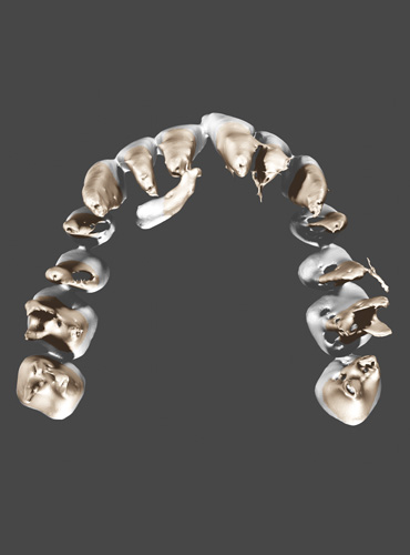
Here is another video of a D3M model on a mobile phone. The screen-captured video depicts bilateral impacted teeth and their position in relation to bone and mandibular canal. The teeth, bone, and canal are separated and viewed as individual rendered models, which help the clinician to plan surgery and the patient understand his situation.
Conclusion
Diagnostic 3D Modeling (D3M) represents a groundbreaking advancement in the field of healthcare, especially in oral radiology and surgery. This innovative technology empowers diagnosticians, clinicians, and surgeons to explore and understand complex pathologies with unprecedented clarity and precision. D3M not only enhances patient education by providing tangible visualizations of their conditions but also aids healthcare professionals in making informed decisions about treatment strategies.
The ability to conduct virtual mock surgeries and design procedures based on an individual’s unique anatomical structures is transformative. Moreover, the recent integration of virtual reality devices further propels D3M into the future, offering an immersive, interactive dimension to medical practice.
As D3M continues to evolve, it promises to redefine the standards of patient care and the capabilities of medical professionals, ultimately leading to better outcomes and improved overall well-being for patients in need of oral health solutions.
- The First Step to Success in Guided Implantology: Dejugadification of the Process - June 28, 2025
- Enlightening Dental Practice: 3D Printed Anatomy Models for Mock Surgeries and Patient Education - September 5, 2023
- Precision and Comfort: 3D Printed Surgical Guides for Implant Placement - September 5, 2023

